

Vitreoretinal Surgery: Retina Health Treatments
Vitreoretinal surgery is a modern approach that addresses the diagnosis, treatment, and surgical management of retinal conditions using advanced technologies. You can continue reading to find essential information about retinal detachment, diabetic retinopathy, macular disorders, and related surgical procedures.
15,000+ Successful Eye Surgeries
Private Insurance Accepted
20+ Years of Experience
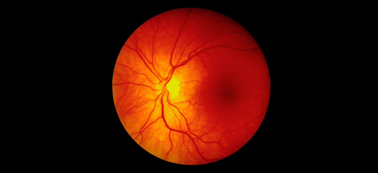
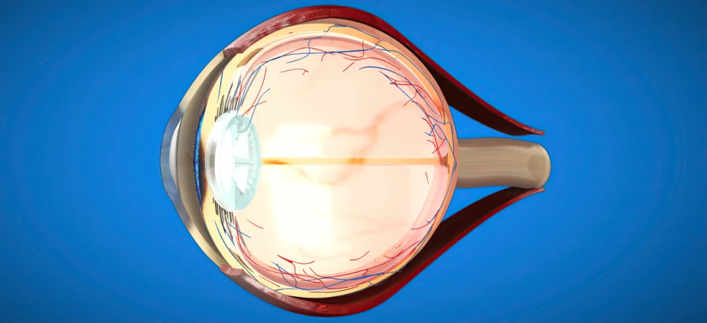
The eye is one of the most important organs for perceiving the world. The retina, which lines the back of the eye and functions much like the film in a camera, is where the visual process takes place. Conditions affecting this delicate layer or the vitreous gel that fills the inside of the eye often require specialized expertise and advanced technology.
Vitreoretinal surgery represents one of the most advanced fields of ophthalmic surgery, aiming to repair these sensitive structures and preserve visual function. In the clinic, modern technologies are used for the diagnosis and management of retinal conditions, with treatment approaches tailored to the specific needs of each case.
What Are the Common Signs of Retinal Problems?
Retinal conditions are typically painless but may present with noticeable changes in visual quality. Common signs may include:
- Floaters: Sudden appearance of dark spots, cobweb-like shapes, or moving specks in the field of vision
- Flashes of Light: Brief flashes resembling lightning or camera flashes, often more noticeable with eye movements
- Curtain-Like Shadow: A dark shadow or curtain appearing from the top, bottom, or side of the visual field
- Distorted Vision: Straight lines (such as door frames or tile edges) appearing wavy or bent
- Central Vision Loss: Blurring or loss of clarity at the point of focus
Conditions Commonly Managed With Vitreoretinal Surgery
Vitreoretinal surgery is an advanced treatment area focused on the retina, one of the most delicate structures of the eye. Both sudden-onset emergencies and slowly progressive conditions that may threaten vision can be addressed using modern surgical techniques.
1. Retinal Detachment
Retinal detachment is a serious ocular condition that often presents with sudden symptoms and requires prompt intervention. Typical signs include flashes of light, floaters, or a curtain-like shadow in the visual field.
This condition occurs when the retina separates from the underlying tissue that supplies it with oxygen and nutrients. Without timely management, retinal function may deteriorate over time.
During vitreoretinal surgery, the retina is repositioned, and supportive techniques such as gas, silicone oil, or laser applications may be used to help secure retinal attachment. The primary aim is to preserve visual function and support a return to daily activities.
2. Diabetic Retinopathy
Diabetes affects not only blood sugar levels but also small blood vessels throughout the body, including those in the eye. Over time, retinal vessels may weaken, leak, bleed, or form abnormal new vessels.
As the condition progresses, bleeding into the eye, traction on the retina, or membrane formation may occur, potentially leading to significant visual impairment.
With vitrectomy surgery:
- Intraocular bleeding can be removed
- Tractional membranes can be carefully peeled using microsurgical instruments
- Retinal anatomy and intraocular pressure can be stabilized
Timely surgical intervention plays an important role in managing diabetes-related retinal damage.
3. Macular Hole
The macula is responsible for the sharpest central vision. Age-related structural changes or traction from the vitreous gel may lead to the formation of a small tear or hole in this area.
Macular hole–related changes may include:
- Difficulty reading, with missing letters or words
- Reduced ability to recognize faces
- Blurring or darkening in the center of vision
This condition does not typically resolve on its own and may progress over time. Surgical techniques are used to relieve traction and support hole closure. Postoperative positioning may be recommended to aid recovery.
4. Epiretinal Membrane (Macular Pucker)
An epiretinal membrane is a thin layer of tissue that can form on the retinal surface over time. By contracting, it may distort the retina and affect image quality.
Common observations include:
- Wavy or distorted vision
- Changes in object shape
- General visual blurring
During vitreoretinal surgery, the membrane is delicately removed using microsurgical techniques. Reducing traction on the retina may lead to gradual visual improvement.
Vitreoretinal Treatment Methods and Current Technologies
Each eye structure and disease progression pattern differs. For this reason, evaluation begins with detailed retinal imaging, high-resolution diagnostic tools, and comprehensive examination. Based on findings, the most appropriate management approach is determined.
Treatment options may include observation, medication, laser therapy, intraocular injections, or advanced vitreoretinal surgery. The goal is always to achieve optimal outcomes with the least necessary intervention.
Pars Plana Vitrectomy (PPV)
Pars plana vitrectomy is one of the most advanced vitreoretinal surgical techniques. Through very small entry points in the white part of the eye, access to the vitreous cavity is achieved with minimal tissue disruption.
How is it performed?
Under high-precision surgical microscopes and imaging systems, the vitreous gel is carefully removed. Retinal membranes are peeled with microsurgical instruments, and retinal tears may be reinforced with laser treatment. The aim is to restore normal retinal anatomy and support visual function.
Modern 23G and 25G systems often allow surgery to be completed without sutures, contributing to faster recovery and improved postoperative comfort.
Intraocular Injections
Not all retinal conditions require surgery. In certain cases—such as macular disease, diabetes-related retinal swelling, or vascular leakage—special medications administered into the eye may be effective.
These procedures are brief, typically performed in an outpatient setting, and aim to reduce retinal swelling, control bleeding, and slow disease progression. With regular follow-up and appropriate treatment planning, visual deterioration may be limited in many cases.
Argon Laser Therapy
Argon laser therapy is a widely used and effective method in retinal care. It is particularly important in managing retinal tears and similar conditions.
Performed in a clinical setting, laser treatment strengthens weak retinal areas by creating a protective barrier. This approach may help prevent progression to retinal detachment and reduce the need for urgent surgery.
The Vitreoretinal Surgery Process
Before surgery, the condition of the eye is carefully evaluated using comprehensive examination and imaging techniques such as OCT. Many vitreoretinal procedures can be performed under local anesthesia, allowing comfort without pain during the operation.
Surgical duration typically ranges from 30 minutes to 1.5 hours, depending on the condition being addressed. Procedures are carried out in a sterile operating environment using advanced equipment and microsurgical tools.
In some cases, a tamponade substance may be placed inside the eye to support retinal healing:
- Gas Tamponade: Absorbs naturally over time; temporary blurred vision is expected, and air travel should be avoided until the gas is fully absorbed.
- Silicone Oil: Does not absorb on its own and may be removed with a minor procedure after healing, usually within several months.
Postoperative care is as important as the surgery itself. Specific head positioning, such as face-down positioning for a limited period, may be recommended to support retinal healing. Mild irritation, blurred vision, or a sensation of heaviness can be part of the normal recovery process. Regular follow-up examinations allow close monitoring of healing progress.