05/Retina and Vitreous Surgery
Retina Surgery: Vitreoretinal Procedures and Techniques
Vitrectomy and vitreoretinal surgery for conditions such as retinal tears and detachment, vitreous hemorrhage, macular holes and epiretinal membranes.
- Examination, surgery and follow-up with the same doctor
- Contracts with 7 private health insurers
- Bağdat Caddesi, Kadıköy


Short answer
Vitreoretinal surgery covers operations on the vitreous gel and the retina at the back of the eye. It is used for conditions such as retinal detachment, vitreous hemorrhage, macular hole and epiretinal membrane. Sudden vision loss, flashes of light or a curtain coming down over your field of vision need urgent assessment.
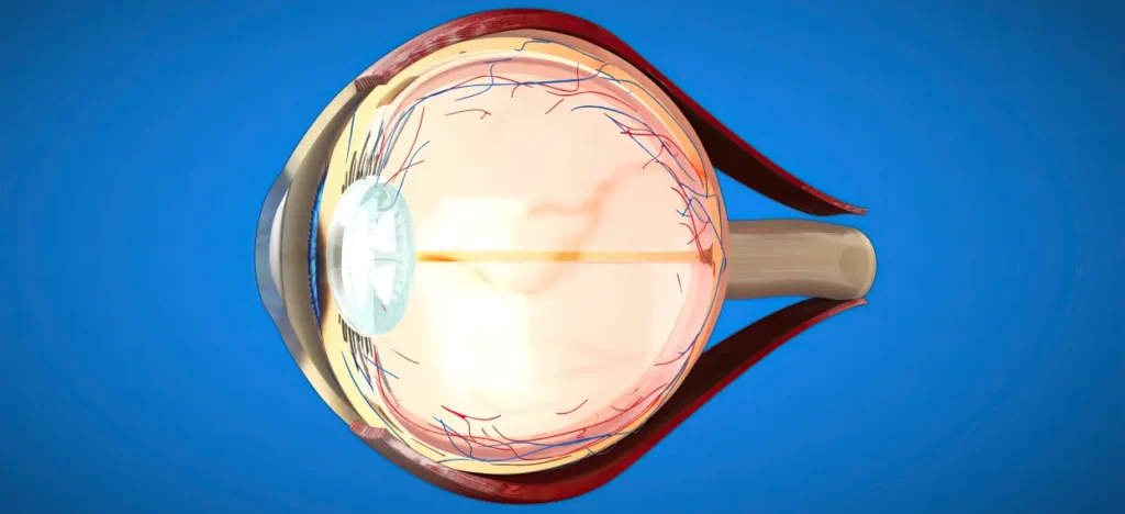
Our eyes are how we take in the world around us. The retina, the layer that lines the back of the eye and works much like the film in a camera, is where vision takes place. Problems in this delicate layer, or in the vitreous gel that fills the inside of the eye, call for specialized expertise and advanced technology.
Vitreoretinal surgery is the field of eye surgery used to repair these delicate structures and preserve vision. Modern technology is used in the diagnosis and treatment of retinal diseases, and every treatment plan is tailored to the individual patient.
What Symptoms Do Retina Problems Cause?
Retinal diseases usually do not cause pain, but they show up as clear changes in the quality of your vision. If you experience any of the following symptoms, you should have an eye exam without delay:
- Floaters: Black dots, cobweb-like specks or fly-like shapes that suddenly appear in front of your eye.
- Flashes of Light: Sudden bursts of light, like lightning or a camera flash (especially if they increase with eye movement).
- A Curtain Over Your Vision: A dark curtain coming down or closing in from one edge of your field of vision (from above, below or the side).
- Distorted Vision: Straight lines (such as door frames or tile lines) looking wavy, broken or crooked.
- Loss of Central Vision: The point you are looking at appearing blurred, or not being able to see it at all.
Main Conditions Treated with Vitreoretinal Surgery
Vitreoretinal surgery is an advanced field of treatment that focuses on the retina, one of the most delicate parts of the eye. It can be used to treat both sudden emergencies and chronic diseases that progress slowly and threaten vision. Below you can read more about the retinal conditions most commonly treated with this type of surgery.
1. Retinal Detachment
Retinal detachment is a serious eye condition that often starts with sudden symptoms and needs prompt treatment. Patients usually notice a dramatic change, which they describe as flashes of light, black floaters or a curtain coming down over their field of vision.
It happens when the retina separates from the layer beneath it that nourishes it. Because the detached retina no longer receives oxygen and nutrients, it gradually stops working. The longer treatment is delayed, the more likely the damage is to become permanent.
During vitreoretinal surgery, the retina is put back into position, and supporting methods (gas, silicone oil, laser) help it stay attached. The aim is both to preserve vision and to help the patient regain their quality of life as soon as possible.
2. Diabetic Retinopathy (Retinal Damage Caused by Diabetes)
Diabetes is not only a matter of blood sugar; over time it affects the small blood vessels throughout the body. The blood vessels of the eye are affected too: they gradually weaken and may begin to leak, bleed or form new but unhealthy vessels.
As the disease progresses, blood can fill the space in front of the retina, vision can drop quickly, or membranes can form on the surface of the retina and pull on it. Situations like these can lead to serious vision loss that disrupts daily life.
With vitrectomy surgery:
- blood inside the eye is cleared,
- membranes pulling on the retina are removed with microsurgical instruments,
- the surface of the retina is restored as far as possible.
Surgery performed at the right time plays an important role in bringing diabetes-related damage to the eye under control.
3. Macular Hole
The macula is the area of the retina that gives us our sharpest vision. Age-related structural changes, or traction from the vitreous (the gel inside the eye), can cause a small tear or hole to form in this area.
People with a macular hole:
- see the middle of words go missing when reading,
- have difficulty recognizing faces,
- notice blurring or darkening at the center of what they look at.
A macular hole does not heal on its own and can grow larger over time.
During surgery, special techniques are used to relieve tension on the retina so that the hole can close, and a short period of face-down positioning after surgery supports healing. With successful treatment, patients can regain much clearer central vision.
4. Epiretinal Membrane (Macular Pucker)
An epiretinal membrane is a thin but significant layer of tissue that gradually forms on the surface of the retina. It adheres to the retina, tightens it almost like a film and distorts the image.
Patients usually begin to:
- see lines as wavy or crooked,
- notice that the shapes of objects look distorted,
- experience noticeable blurring of vision.
During vitreoretinal surgery, this membrane is peeled away with great care. The procedure is done with microsurgical techniques that require millimeter-level precision. Once the membrane is removed, the pull on the retina is released and vision can improve noticeably over time.
Vitreoretinal Surgery Methods and Current Technology
Every person’s eye anatomy, the cause of their symptoms and the speed at which the disease progresses are different. That is why treatment begins with a fully individual assessment rather than a single standard approach. Detailed retinal scans, high-resolution imaging and advanced diagnostic devices used at the first examination help identify the source of the problem clearly.
The most suitable treatment is then chosen according to the type of disease and the needs of the eye. Sometimes this means monitoring and medication alone; in other cases laser treatment, eye injections or advanced vitreoretinal surgical techniques may be needed. The aim is always to achieve the result the eye needs with as little intervention as possible.
Pars Plana Vitrectomy (PPV)
Pars plana vitrectomy is a cornerstone of modern vitreoretinal surgery. The surgeon reaches the inside of the eye through openings as fine as the tip of a needle, made in the white of the eye. This allows high precision with minimal contact with the tissues.
How is it performed?
Using a special operating microscope and high-resolution viewing systems, the gel that naturally fills the eye (the vitreous) is carefully removed. Membranes on the retina are then peeled away with fine micro-instruments, and any tears are reinforced with laser. The aim is to restore the normal anatomy of the retina and to improve vision as much as possible.
With 23-gauge (23G) and 25-gauge (25G) systems, most vitrectomies can be completed without stitches. This not only speeds up recovery after surgery but also makes the return to daily life much more comfortable.
Eye Injections
Not every retinal disease needs surgery. In some conditions, particularly macular degeneration, diabetic macular edema or blood vessel problems that cause leakage inside the eye, special medicines injected into the eye can be a very effective treatment.
The procedure is quick, is carried out under sterile conditions and can usually be done without disrupting your daily life. Injections aim to reduce swelling in the retina, control bleeding and halt the progression of the disease. With regular follow-up and careful planning of the medication, vision loss can be prevented in many patients.
Argon Laser Treatment
Argon laser is a commonly used, preventive and effective treatment for retinal diseases. It is especially important in conditions that can develop suddenly, such as retinal tears. The treatment is done in the clinic, takes a short time and strengthens weak areas of the retina.
The laser spots placed around the tear act as a barrier and help prevent the tear from turning into a retinal detachment. In many patients, this stabilizes the retina without the need for emergency surgery.
The Vitreoretinal Surgery Process
Before the day of surgery, the condition of your eye is assessed in detail. A comprehensive eye exam may be carried out, and the structure of the retina may be examined with imaging tests such as OCT. Most vitreoretinal operations can be performed under local anesthesia. This means the eye is fully numbed: you can stay awake throughout the operation, and pain is usually not felt. Patients who feel excited, tense or anxious may be given a mild sedative so that the process is more comfortable.
Depending on the condition being treated, vitreoretinal surgery usually takes between 30 minutes and 1.5 hours.
The operation is performed in a fully sterile operating room with advanced equipment. The necessary repairs are made inside the eye with millimeter-scale instruments. During this stage, all you need to do is stay calm and follow the team’s instructions.
In some retinal operations, a tamponade agent may be placed inside the eye to support the healing of the retina. This helps keep the retina in the desired position.
- Gas Tamponade:
The gas stays in the eye for a certain period and is gradually absorbed on its own. Your vision may be blurred while the gas is present; this is completely normal. You must also not travel by air until the gas has completely disappeared. - Silicone Oil:
Unlike gas, silicone oil does not disappear on its own. It is usually removed with a minor procedure once healing is complete, generally within 3–6 months. Silicone oil may be preferred when longer-lasting support is needed.
The period after surgery is as important for the success of treatment as the surgery itself. If gas or silicone oil has been placed in the eye, your doctor may recommend a specific head position for a certain period.
This is often a “face-down position” and usually lasts 3–7 days. This position helps the tamponade stay in full contact with the retina and supports healing.
Mild stinging, blurred vision or a feeling of heaviness in the eye can be normal during recovery. Regular check-ups allow your recovery to be monitored closely.
The information on this page is for general information only and does not replace diagnosis or treatment. In any surgical or interventional procedure, results vary from person to person. Please get a detailed opinion from your doctor before any procedure.

Frequently asked questions
Retina Surgery: frequently asked questions
If you have another question, you can message us on WhatsApp.
Is retinal detachment an emergency?
Yes. Sudden flashes of light, a sudden increase in floaters and a curtain coming down over your field of vision can be signs of retinal detachment; you should have an eye exam without delay.
Do I need to lie face down after surgery?
If gas has been placed in the eye, a specific head position may be recommended for a certain period to help the retina stay in place. How long and in which position will be explained to you separately, depending on the type of surgery.

Your doctor
Op. Dr. Murat Ün
He graduated from Trakya University Faculty of Medicine and completed his ophthalmology residency at Fatih Sultan Mehmet Training and Research Hospital. From 2004 to 2016 he worked at Dünyagöz Hospital in Ataköy, then at Medical Park and Batıgöz. Since 2021 he has been seeing patients at his own practice on Bağdat Caddesi.
- Education
Trakya University Faculty of Medicine
- Residency
Fatih Sultan Mehmet Training and Research Hospital, Ophthalmology
- 2001–2004
Yaşar Okuyan SSK Hospital
- 2004–2016
Dünyagöz Hospital, Ataköy
- 2016–2018
Medical Park Hospital
- 2018–2021
Batıgöz Health Group, Altunizade
- 2021 →
Own practice · Bağdat Caddesi, Kadıköy
Contracted insurers
Examination with your private health insurance
Before your appointment we confirm together what your policy covers for Retina Surgery. Coverage and any co-payment depend on your insurer’s approval.
- 01
 Allianz SigortaContracted
 - 02
 Anadolu SigortaContracted
 - 03
 SencardContracted
 - 04
 Türkiye SigortaContracted
 - 05
 MAPFRE SigortaContracted
 - 06
 Medisa SigortaContracted
 - 07
 AXA SigortaContracted
 - Insurer not on the list?Share your policy details and we will look at reimbursement options together.Ask on WhatsApp